Visualizing Protein-Ligand Unpaired Interactions
Problem
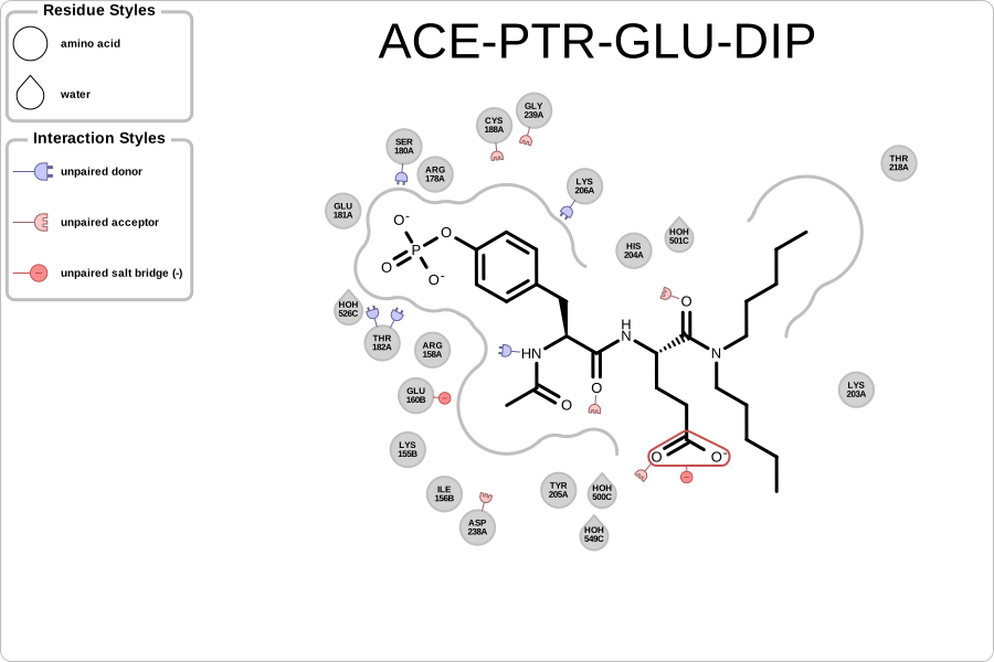
You want to visualize protein-ligand unpaired and clash interactions. See example in Figure 1.
Figure 1. Example of visualizing protein-ligand unpaired and clash interactions (PDB: 1NQ2))
Ingredients
|
Difficulty level
🌶️ 🌶️
Download
Source Code
unpairedmap2img
#!/usr/bin/env python3
# (C) 2023 Cadence Design Systems, Inc. (Cadence)
# All rights reserved.
# TERMS FOR USE OF SAMPLE CODE The software below ("Sample Code") is
# provided to current licensees or subscribers of Cadence products or
# SaaS offerings (each a "Customer").
# Customer is hereby permitted to use, copy, and modify the Sample Code,
# subject to these terms. Cadence claims no rights to Customer's
# modifications. Modification of Sample Code is at Customer's sole and
# exclusive risk. Sample Code may require Customer to have a then
# current license or subscription to the applicable Cadence offering.
# THE SAMPLE CODE IS PROVIDED "AS IS", WITHOUT WARRANTY OF ANY KIND,
# EXPRESS OR IMPLIED.  OPENEYE DISCLAIMS ALL WARRANTIES, INCLUDING, BUT
# NOT LIMITED TO, WARRANTIES OF MERCHANTABILITY, FITNESS FOR A
# PARTICULAR PURPOSE AND NONINFRINGEMENT. In no event shall Cadence be
# liable for any damages or liability in connection with the Sample Code
# or its use.

"""Depict the unpaired and clash interactions of an active site."""

import argparse
import io
import os
import sys
from pathlib import Path

from openeye import oechem, oedepict, oegrapheme
from PIL import Image
from rich_argparse import HelpPreviewAction, RichHelpFormatter

__SCRIPT_NAME__ = Path(__file__).absolute().stem
__SCRIPT_DESC__ = "Depict the unpaired and clash interactions of an active site."
__SCRIPT_TOOLKITS__ = ["oechem", "oedepict", "oegrapheme"]
__SCRIPT_CATEGORIES__ = ["visualization", "ligand-protein interactions"]


def parse_options() -> argparse.Namespace:
    """Set up command line options."""
    parser = argparse.ArgumentParser(
        add_help=True,
        formatter_class=RichHelpFormatter,
        description="[yellow]" + __SCRIPT_DESC__ + "[/yellow]",
    )
    parser.add_argument("--help-image", action=HelpPreviewAction)

    # input options
    input_group = parser.add_argument_group("Input ligand-protein complex")
    exclusive_input_group = input_group.add_mutually_exclusive_group(required=True)
    exclusive_input_group.add_argument(
        "--complex",
        type=str,
        required=False,
        metavar="PDB-FILE",
        help="input PDB file of the ligand-protein complex",
    )
    exclusive_input_group.add_argument(
        "--design-unit",
        "--du",
        type=str,
        metavar="DU-FILE",
        help="input design unit file",
    )
    image_group = parser.add_argument_group("Image options")
    image_group.add_argument(
        "--image",
        type=str,
        required=False,
        metavar="IMAGE-FILE",
        help="output image file (PNG, SVG) (required: %(required)s) -- if no output is provided the image will be displayed on the  screen",
    )
    image_group.add_argument(
        "--width",
        type=int,
        default=900,
        help="width of output image (default: %(default)s)",
    )
    image_group.add_argument(
        "--height",
        type=int,
        default=600,
        help="height of output image (default: %(default)s)",
    )
    image_group.add_argument(
        "--interactive-legend",
        default=False,
        action="store_true",
        help="visualize legend on mouse hover (SVG-only feature) (default: %(default)s)",
    )
    return parser.parse_args()


def main() -> int:
    """Depict the unpaired and clash interactions of an active site."""
    args = parse_options()

    _check_image_file(args)

    if args.complex:
        protein, ligand = get_protein_and_ligand_from_pdb(args.complex)
    elif args.design_unit:
        protein, ligand = get_protein_and_ligand_from_design_unit(args.design_unit)
    else:
        oechem.OEThrow.Fatal("Invalid input option!")

    # depict unpaired interaction map
    image = oedepict.OEImage(args.width, args.height)

    cell_width, cell_height = args.width, args.height
    if not args.interactive_legend:
        cell_width = cell_width * 0.8

    opts = oegrapheme.OE2DActiveSiteDisplayOptions(cell_width, cell_height)
    opts.SetRenderInteractiveLegend(args.interactive_legend)

    if args.interactive_legend:
        depict_unpaired_map(image, protein, ligand, opts)
    else:
        main_frame = oedepict.OEImageFrame(
            image,
            args.width * 0.80,
            args.height,
            oedepict.OE2DPoint(args.width * 0.2, 0.0),
        )
        legend_frame = oedepict.OEImageFrame(
            image,
            args.width * 0.20,
            args.height,
            oedepict.OE2DPoint(args.width * 0.0, 0.0),
        )
        depict_unpaired_map(main_frame, protein, ligand, opts, legend_frame)

    if (
        args.image
        and Path(args.image).suffix[1:].lower() == "svg"
        and args.interactive_legend
    ):
        icon_scale = 0.5
        oedepict.OEAddInteractiveIcon(
            image, oedepict.OEIconLocation_TopRight, icon_scale
        )
    oedepict.OEDrawCurvedBorder(image, oedepict.OELightGreyPen, 10.0)

    if args.image:
        oedepict.OEWriteImage(args.image, image)
    else:
        _img = Image.open(io.BytesIO(oedepict.OEWriteImageToBytes("png", image)))
        _img.show()

    return os.EX_OK


def depict_unpaired_map(
    image: oedepict.OEImageBase,
    protein: oechem.OEMolBase,
    ligand: oechem.OEMolBase,
    depict_options: oegrapheme.OE2DActiveSiteDisplayOptions,
    legend_frame: oedepict.OEImageBase | None = None,
) -> None:
    """Depict unpaired interaction map."""
    # perceive interactions
    active_site = oechem.OEInteractionHintContainer(protein, ligand)
    if not active_site.IsValid():
        oechem.OEThrow.Fatal("Cannot initialize active site!")
    active_site.SetTitle(ligand.GetTitle())

    oechem.OEPerceiveInteractionHints(active_site)

    # depiction

    oegrapheme.OEPrepareActiveSiteDepiction(active_site)
    active_site_disp = oegrapheme.OE2DActiveSiteDisplay(active_site, depict_options)
    oegrapheme.OERenderUnpairedInteractionMap(image, active_site_disp)

    if legend_frame:
        legend_options = oegrapheme.OE2DActiveSiteLegendDisplayOptions(12, 1)
        oegrapheme.OEDrawUnpairedInteractionMapLegend(
            legend_frame, active_site_disp, legend_options
        )


def get_protein_and_ligand_from_pdb(
    pdb_filename: str,
) -> tuple[oechem.OEMolBase, oechem.OEMolBase]:
    """Read protein and and ligand from from pdb/cif file."""
    ifs = oechem.oemolistream()
    if not ifs.open(pdb_filename):
        oechem.OEThrow.Fatal(f"Unable to open {pdb_filename} for reading")

    complex_mol = oechem.OEGraphMol()
    if not oechem.OEReadMolecule(ifs, complex_mol):
        oechem.OEThrow.Fatal(f"Unable to read complex from {pdb_filename}")

    if not oechem.OEHasResidues(complex_mol):
        oechem.OEPerceiveResidues(complex_mol, oechem.OEPreserveResInfo_All)

    # separate ligand and protein
    split_opts = oechem.OESplitMolComplexOptions()
    ligand = oechem.OEGraphMol()
    protein = oechem.OEGraphMol()
    water = oechem.OEGraphMol()
    other = oechem.OEGraphMol()

    split_opts.SetProteinFilter(
        oechem.OEOrRoleSet(split_opts.GetProteinFilter(), split_opts.GetWaterFilter())
    )
    split_opts.SetWaterFilter(
        oechem.OEMolComplexFilterFactory(oechem.OEMolComplexFilterCategory_Nothing)
    )

    oechem.OESplitMolComplex(ligand, protein, water, other, complex_mol, split_opts)

    if ligand.NumAtoms() == 0:
        oechem.OEThrow.Fatal("Cannot separate complex!")

    return protein, ligand


def get_protein_and_ligand_from_design_unit(
    filename: str,
) -> tuple[oechem.OEMolBase, oechem.OEMolBase]:
    """Read protein and and ligand from from design unit file."""
    du = oechem.OEDesignUnit()
    if not oechem.OEIsReadableDesignUnit(filename) or not oechem.OEReadDesignUnit(
        filename, du
    ):
        oechem.OEThrow.Fatal("Cannot read design unit.")

    protein = oechem.OEGraphMol()
    if not du.GetComponents(protein, oechem.OEDesignUnitComponents_TargetComplex):
        oechem.OEThrow.Fatal("Could not extract protein from the design unit.")

    ligand = oechem.OEGraphMol()
    if not du.GetLigand(ligand):
        oechem.OEThrow.Fatal("Could not extract ligand from the design unit.")

    return (protein, ligand)


def _check_image_file(args: argparse.Namespace) -> None:
    # script will terminate if there is some issues
    if not args.image:
        # image will be displayed on the screen
        return
    ext = Path(args.image).suffix[1:].upper()
    if not oedepict.OEIsRegisteredImageFile(ext):
        oechem.OEThrow.Fatal("Unknown image output type!")

    ofs = oechem.oeofstream()
    if not ofs.open(args.image):
        oechem.OEThrow.Fatal("Cannot open output image file!")


setattr(main, "__SCRIPT_NAME__", __SCRIPT_NAME__)
setattr(main, "__SCRIPT_DESC__", __SCRIPT_DESC__)
setattr(main, "__SCRIPT_TOOLKITS__", __SCRIPT_TOOLKITS__)
setattr(main, "__SCRIPT_CATEGORIES__", __SCRIPT_CATEGORIES__)

if __name__ == "__main__":
    sys.exit(main())
Solution
The depict_unpairedmap illustrates how simple it is to generate these images.
OEInteractionHintContainer object is constructed that stores information about possible interactions between the ligand and the protein.
The interactions are perceived by calling the OEPerceiveInteractionHints function.
The active site is then prepared for 2D depiction by invoking the OEPrepareActiveSiteDepiction function.
When the OE2DActiveSiteDisplay object is constructed, residues are positioned around the ligand close to those atoms which they are interacting with.
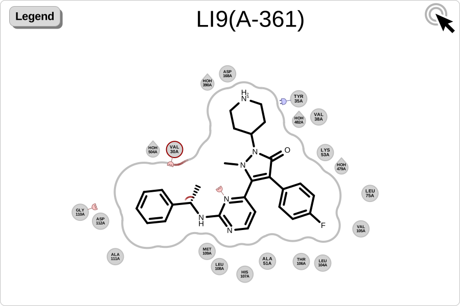
The OERenderUnpairedInteractionMap function generates an image that displays the clash and unpaired interactions detected in the ligand and in nearby residues.
The legend associated with the unpaired map is rendered by invoking the OEDrawUnpairedInteractionMapLegend function.
def depict_unpaired_map(
    image: oedepict.OEImageBase,
    protein: oechem.OEMolBase,
    ligand: oechem.OEMolBase,
    depict_options: oegrapheme.OE2DActiveSiteDisplayOptions,
    legend_frame: oedepict.OEImageBase | None = None,
) -> None:
    """Depict unpaired interaction map."""
    # perceive interactions
    active_site = oechem.OEInteractionHintContainer(protein, ligand)
    if not active_site.IsValid():
        oechem.OEThrow.Fatal("Cannot initialize active site!")
    active_site.SetTitle(ligand.GetTitle())

    oechem.OEPerceiveInteractionHints(active_site)

    # depiction

    oegrapheme.OEPrepareActiveSiteDepiction(active_site)
    active_site_disp = oegrapheme.OE2DActiveSiteDisplay(active_site, depict_options)
    oegrapheme.OERenderUnpairedInteractionMap(image, active_site_disp)

    if legend_frame:
        legend_options = oegrapheme.OE2DActiveSiteLegendDisplayOptions(12, 1)
        oegrapheme.OEDrawUnpairedInteractionMapLegend(
            legend_frame, active_site_disp, legend_options
        )
Usage
See Download section to download the script.
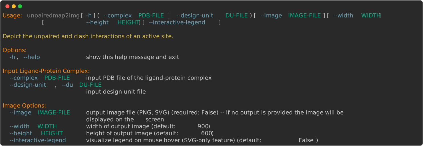
> unpairedmap2img --help
Visualizing 1YWR_DU_0.oedu design unit of 1YWR.
> unpairedmap2img --design-unit 1YWR_DU_0.oedu --interactive-legend --image image.svg
Discussion
Interaction Perception
Currently the OEPerceiveInteractionHints function perceives the following interaction types:
Table 1. Interaction types currently available in OEChem TK name
corresponding interaction class
corresponding interaction type namespace
cation-pi
chelator
clash
None
contact
None
covalent
None
halogen bond
hydrogen bond
salt-bridge
stacking (T and Pi)
The default geometric parameters used by the OEPerceiveInteractionHints function have been set based on literature data ([Kumar-2002], [Cavallo-2016], [Bissantz-2010], and [Marcou-2007] ). The interaction parameters can be customized by using the OEPerceiveInteractionOptions class.
Unpaired Interaction Depiction
The OERenderUnpairedInteractionMap function currently visualizes the following interactions detected by the OEPerceiveInteractionHints function.
Atom clash interaction
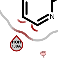
When visualizing protein-ligand atom clashes, a red outline of a residue circle indicates that there are one or more atoms in that residue which are too close to some ligand atom(s). Clashing ligand atoms are marked with a red arc that is directed towards the corresponding clashing residue. The red shading on the grey line representing the shape of the pocket is used to identify atom clashes easily.
Table 2. Examples of visualizing atom clash(es) 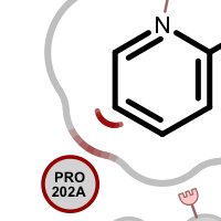
See also
OEClashInteractionHint class in the OEChem TK manual
Unpaired types of the hydrogen bonding interaction
An unpaired hydrogen bond interaction is detected:
if there is no ligand/protein acceptor atom that could interact with a protein/ligand donor atom
if there is no ligand/protein donor atom that could interact with a protein/ligand acceptor atom
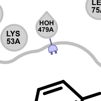
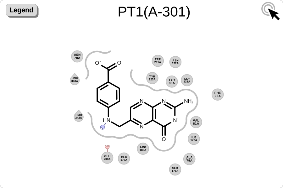
Different linker types are used to mark unpaired acceptor and donor hydrogen bond interactions. Please note that since these interactions are unpaired, the direction of the linkers has no real spatial meaning. Ligand linkers are directed away from the ligand, while protein linkers are directed towards the ligand.
Table 3. Examples of visualizing unpaired hydrogen bond interactions 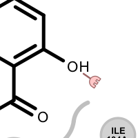
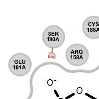
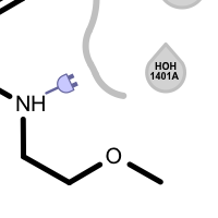
unpaired ligand acceptor
unpaired protein acceptor
unpaired ligand donor
unpaired protein donor
See also
OEHBondInteractionHint class in the OEChem TK manual
OEHBondInteractionHintType namespace in the OEChem TK manual
Clash types of the hydrogen bonding interaction
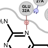
A hydrogen bond clash interaction is detected:
if an acceptor ligand atom interacts with an acceptor protein atom
if a donor ligand atom interacts with an donor protein atom
Table 4. Examples of visualizing hydrogen bond clash interactions 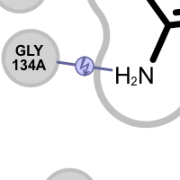
donor-donor clash
acceptor-acceptor clash
See also
OEHBondInteractionHint class in the OEChem TK manual
OEHBondInteractionHintType namespace in the OEChem TK manual
Unpaired types of the salt-bridge interaction
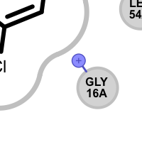
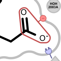
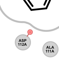
An unpaired salt bridge interaction is detected if there is a positively / negatively charged functional group either in the ligand or in nearby protein without a matching negatively / positively charged functional group, respectively.
Different linker types are used to mark unpaired positive and negative salt-bridge interactions. Please note that since these interactions are unpaired, the direction of the linkers has no real spatial meaning. Ligand linkers are directed away from the ligand, while protein linkers are directed towards the ligand.
Table 5. Examples of visualizing unpaired salt-bridge interactions 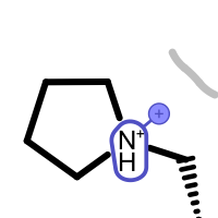
unpaired ligand positive
unpaired protein positive
unpaired ligand negative
unpaired protein negative
See also
OESaltBridgeInteractionHint class in the OEChem TK manual
OESaltBridgeInteractionHintType namespace in the OEChem TK manual
Hydrogen Position Optimization
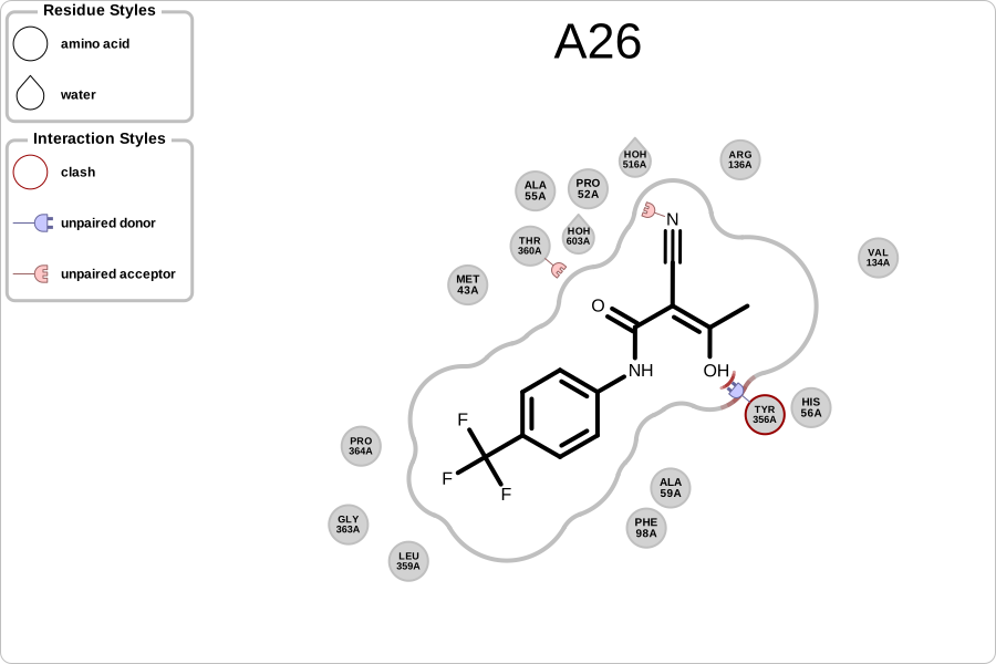
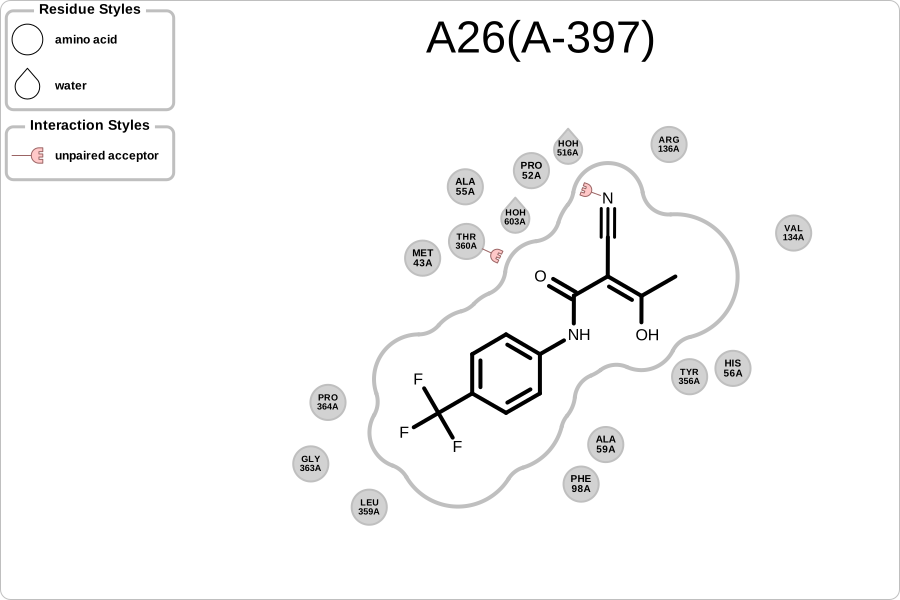
Since interaction perception depends on the position of hydrogens, it is highly recommended to optimize those positions prior to perceiving the interactions. The two images below reveal the effect of optimizing the hydrogen bond network in a protein-ligand complex: fewer atom clashes and fewer unpaired hydrogen bond interactions.
|
|
original complex |
design unit |
See also
OEPlaceHydrogens function in the OEChem TK manual
Protein Preparation chapter in the OEChem TK manual
Creating OEDesignUnits from a PDB file chapter in the Spruce TK manual
Unpaired Map vs Active Site Interaction Map
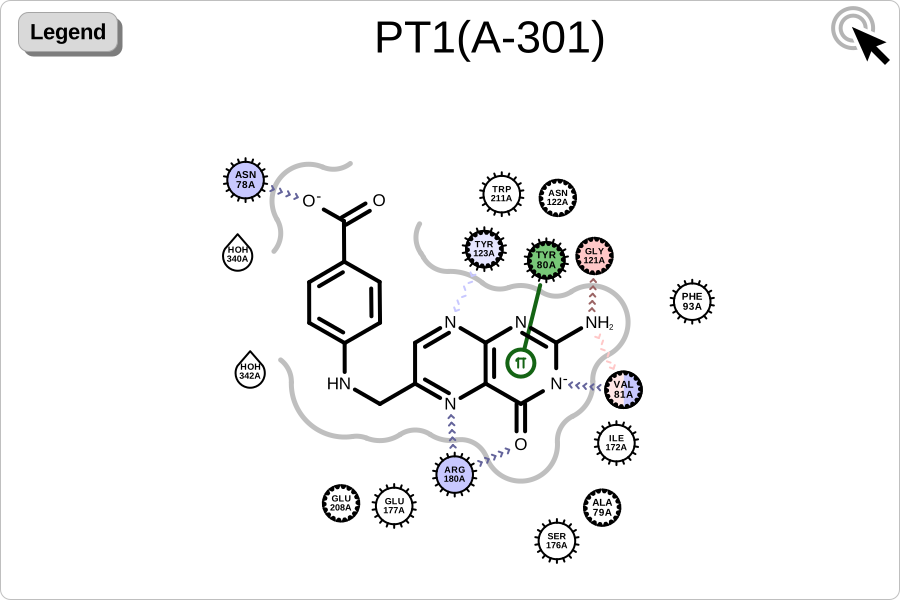
An unpaired interaction map provides a complementary view to the more common active site interaction map. While the interaction map (on the right) depicts interactions between the ligand and protein, the unpaired map (on the left) illustrates interactions that could contribute to binding but are not formed in the complex. Together, these two maps of the protein-ligand binding site provide insights into protein-ligand interactions and communicate complex 3D structural results to medicinal chemists in a directly actionable way.
|
|
unpaired interaction map of |
active site interaction map of |
See also in OEChem TK manual
Theory
Biopolymers chapter
Protein Preparation chapter
API
OEPerceiveInteractionHints function
OEPlaceHydrogens function
See also in OEDepict TK manual
Theory
Molecule Depiction chapter
API
OEImage class
See also in GraphemeTM TK manual
API
OE2DActiveSiteDisplay class
OEDrawUnpairedInteractionMapLegend function
OEPrepareActiveSiteDepiction function
OERenderUnpairedInteractionMap function